Basics of Biochemistry Principles of Exercise
Table of Contents
- Energy Systems and ATP Production
- Glucose Biochemistry
- Substrate Utilization and Hormonal Control
- Lactate Metabolism and the Lactate Threshold
- Fibers, Adaptations, Recovery and Clinical Applications
1 Energy Systems and ATP Production
Learning objectives:
- The three energy systems (ATP-PCr, glycolytic, oxidative) by time-course, ATP rate and capacity
- Why effort duration selects the dominant pathway, and how aerobic and anaerobic ATP yield from glucose differ
- The concept of first and second metabolic thresholds
1.1 Introduction to Exercise Biochemistry
Exercise biochemistry examines the molecular and metabolic changes that occur during physical activity. Understanding these principles is essential for optimising athletic performance, designing effective training programmes, and comprehending how exercise promotes health and prevents disease (Brooks et al. 2005).
At the cellular level, exercise initiates a cascade of biochemical events involving energy metabolism, substrate utilisation, hormone regulation, and adaptive signalling pathways. These processes are highly coordinated and depend on exercise intensity, duration, and the individual’s training status (Hawley et al. 2014).
1.2 The ATP-PCr System (Phosphagen System)
The most immediate energy source for muscle contraction is adenosine triphosphate (ATP). However, intramuscular ATP stores are limited and can sustain maximal exercise for only 2–3 seconds. The phosphocreatine (PCr) system provides rapid ATP regeneration through the creatine kinase reaction (Greenhaff 2001):
PCr + ADP + H⁺ → ATP + Creatine
This system is crucial for high-intensity, short-duration activities (5–10 seconds) such as sprinting, jumping, or powerlifting. Complete PCr restoration requires 3–5 minutes of rest (Harris et al. 1976).
1.3 Glycolytic System (Anaerobic Glycolysis)
When exercise extends beyond 10 seconds, the glycolytic system becomes the predominant ATP source. This pathway breaks down glucose or glycogen without requiring oxygen, producing pyruvate and lactate as end products (Robergs et al. 2004). Key features:
- Net ATP yield: 2–3 ATP per glucose molecule (depending on glucose source)
- Peak power output: 30–90 seconds
- Produces hydrogen ions (H⁺) that contribute to metabolic acidosis
- Critical for activities like the 400 m sprint, 100 m swim, or resistance training
1.4 Oxidative System (Aerobic Metabolism)
The oxidative system provides sustainable ATP production through complete oxidation of carbohydrates and fats in the mitochondria, via three main processes: pyruvate oxidation, the citric acid cycle (Krebs cycle), and the electron transport chain (Holloszy & Coyle 1984).
ATP yields from complete oxidation are substantially higher than anaerobic pathways: approximately 32–36 ATP per glucose molecule and over 100 ATP per palmitate (16-carbon fatty acid). This efficiency makes the oxidative system ideal for prolonged exercise lasting more than 2–3 minutes (Berg et al. 2002).
1.5 Comparison of Energy Systems
The three energy systems compared by the two properties that matter in practice: how fast they resynthesise ATP, and how long they can keep it up. ATP–PCr delivers the highest rate but is spent within 5–10 s, glycolysis peaks over 30–90 s, and oxidative phosphorylation sustains a moderate rate for effectively unlimited duration. Note the logarithmic time axis.
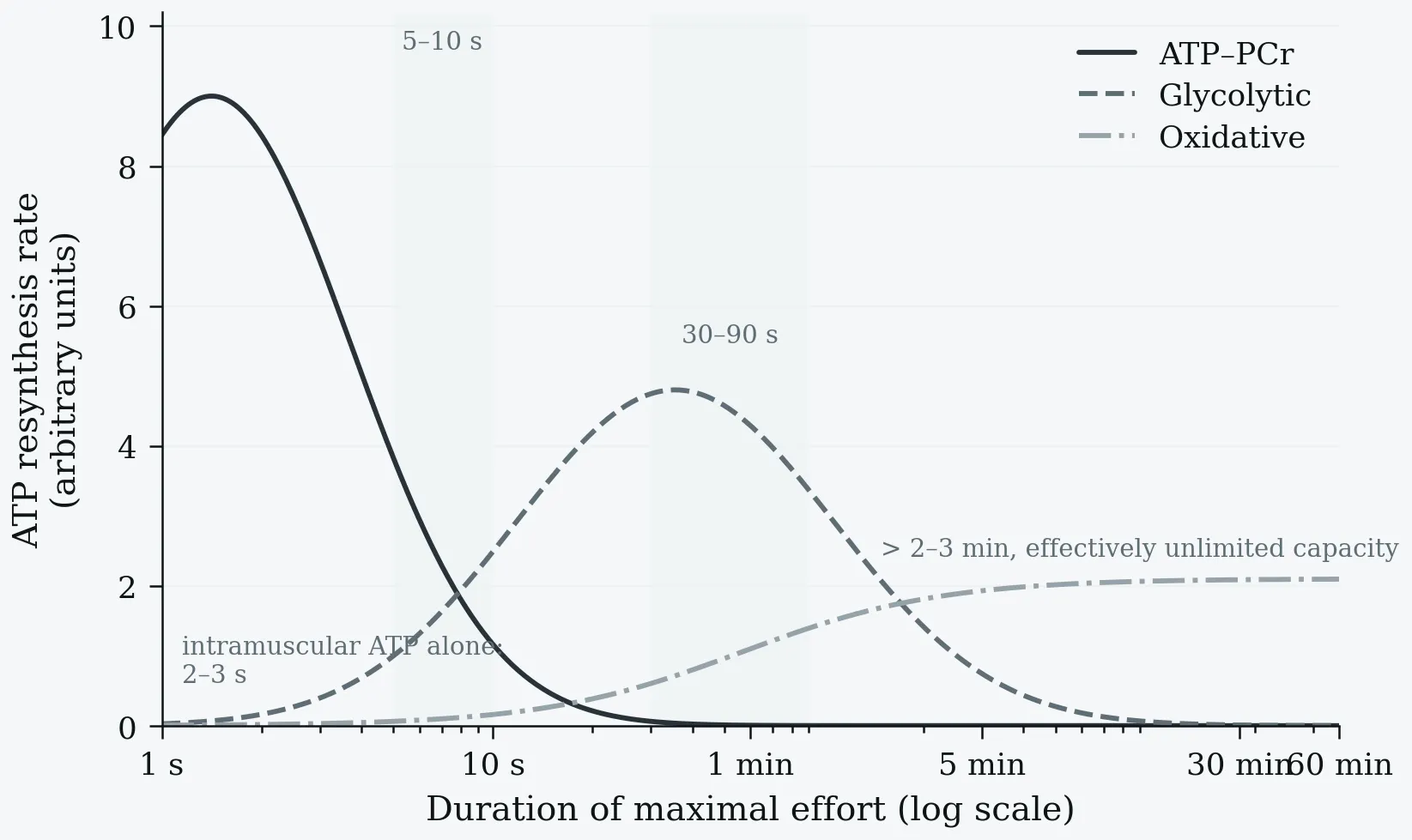
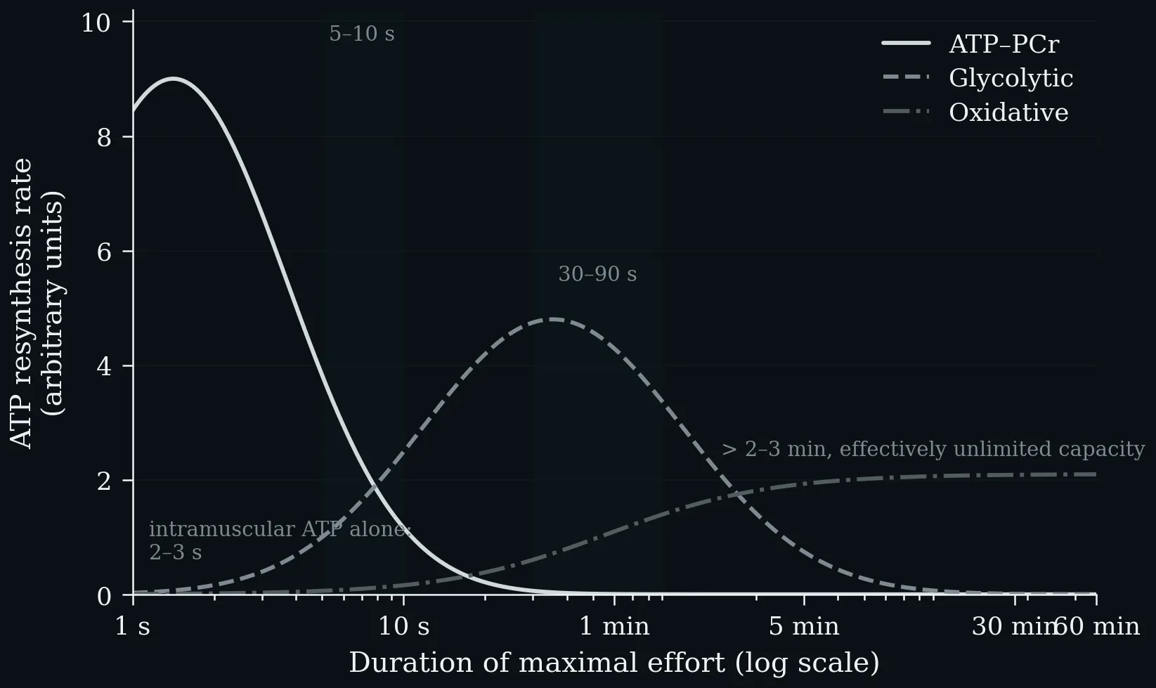
The three energy systems compared by the two properties that matter in practice: how fast they resynthesise ATP, and how long they can keep it up. ATP–PCr delivers the highest rate but is spent within 5–10 s, glycolysis peaks over 30–90 s, and oxidative phosphorylation sustains a moderate rate for effectively unlimited duration. Note the logarithmic time axis.
After uptake into the cell, glucose is phosphorylated and passes through glycolysis down to pyruvate. Under anaerobic conditions, pyruvate is converted to lactate to regenerate the NAD⁺ required for glycolysis, and lactate is typically released by the cells (2 mol ATP per mol glucose). Under aerobic conditions, pyruvate enters the mitochondria and is converted to acetyl-CoA by oxidative decarboxylation, feeding the citrate cycle; it is broken down to CO₂ and the reduction equivalents NADH/H⁺ and FADH₂, whose oxygen-dependent reoxidation yields water and is coupled to ATP synthesis from ADP and Pᵢ, giving ≈ 30 mol ATP per mol glucose.
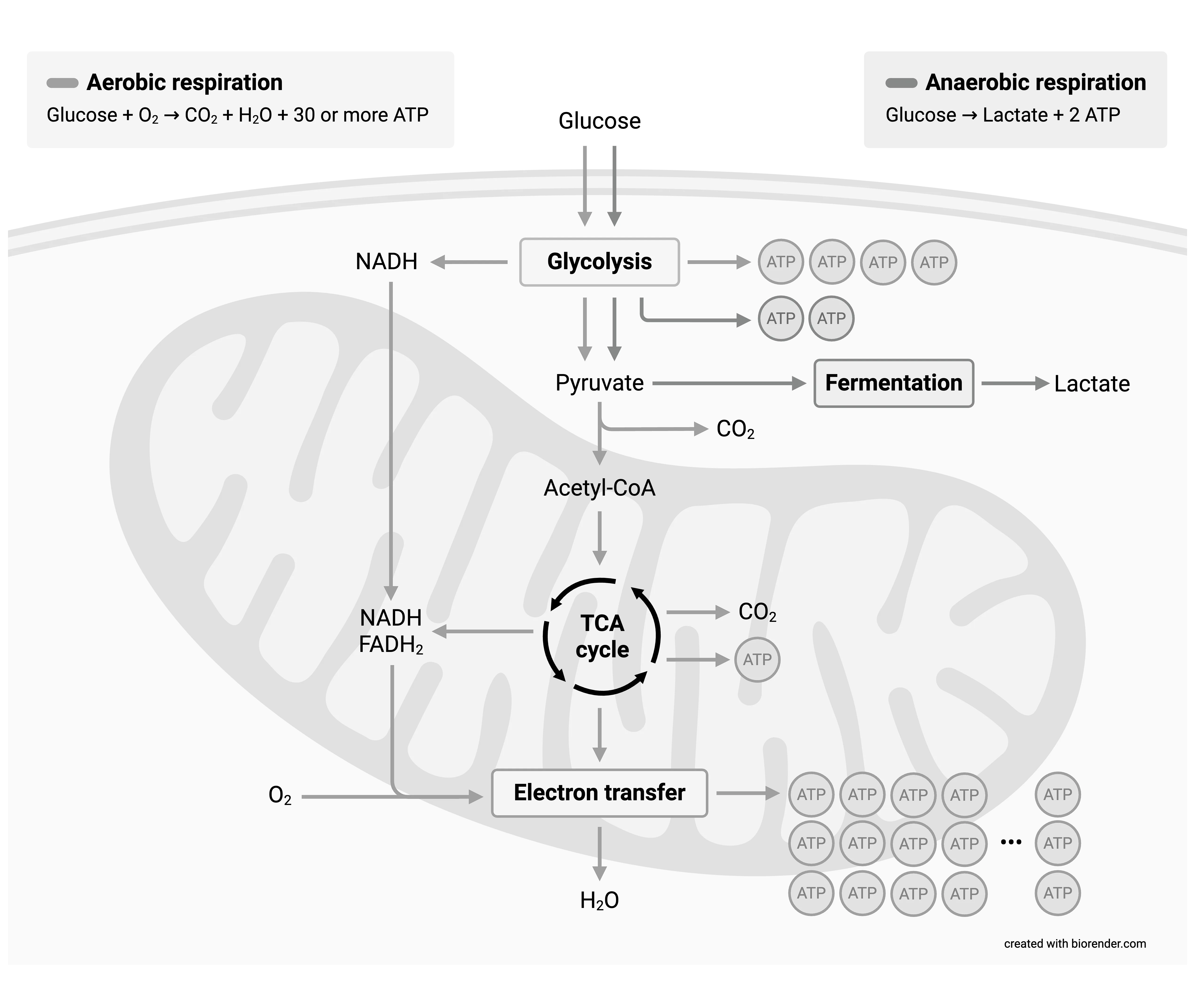
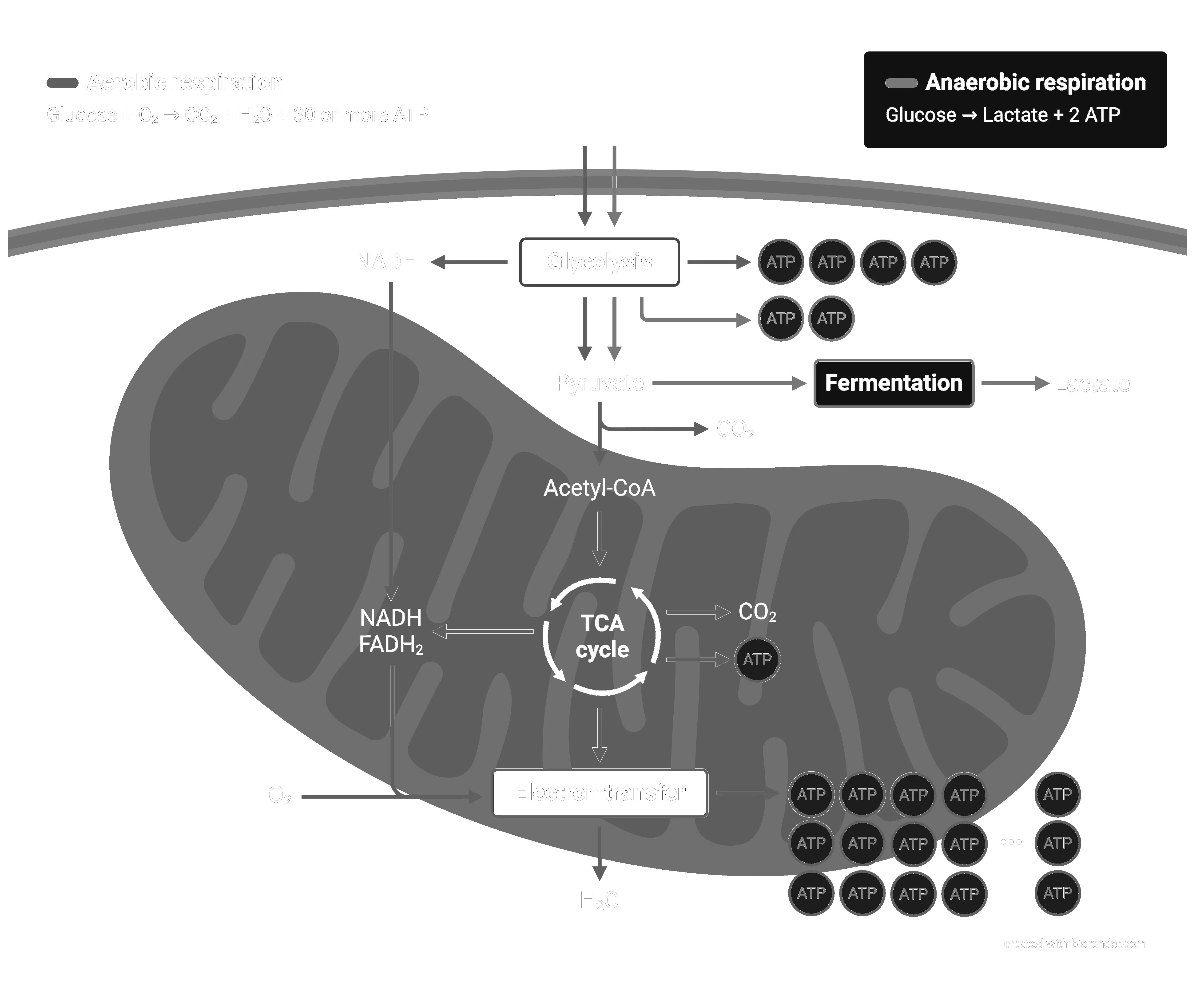
After uptake into the cell, glucose is phosphorylated and passes through glycolysis down to pyruvate. Under anaerobic conditions, pyruvate is converted to lactate to regenerate the NAD⁺ required for glycolysis, and lactate is typically released by the cells (2 mol ATP per mol glucose). Under aerobic conditions, pyruvate enters the mitochondria and is converted to acetyl-CoA by oxidative decarboxylation, feeding the citrate cycle; it is broken down to CO₂ and the reduction equivalents NADH/H⁺ and FADH₂, whose oxygen-dependent reoxidation yields water and is coupled to ATP synthesis from ADP and Pᵢ, giving ≈ 30 mol ATP per mol glucose.
1.6 Common Metabolic Thresholds
Metabolic threshold: an exercise intensity that results in an apparent change in metabolism (usually reflected by changes in oxygen uptake, carbon dioxide production, ventilation, or metabolites in muscle and blood). The first detectable change is often termed the first metabolic threshold, the second detectable change the second metabolic threshold (Bishop et al. 2025).
One-Minute-Paper Topics
A One-Minute-Paper (OMP) is a short, focused prompt that students answer in ~60 seconds at the end of a session to consolidate learning, surface misconceptions, and provide formative feedback. When answering, be concise, specific, and use terminology from today’s session.
- Compare the phosphagen, glycolytic, and oxidative ATP systems by time course and yield.
- Why does the phosphagen system dominate in efforts shorter than ~10 seconds?
- Outline glycolysis in three steps and identify its net ATP yield.
- Why is the aerobic ATP yield from glucose roughly 15× the anaerobic yield, and what limits the anaerobic route?
- Define the first and second metabolic threshold and name one measurable variable that reveals each.
Key References
- Berg JM, Tymoczko JL, Stryer L. Biochemistry, 5th edn. W.H. Freeman and Company, 2002.
- Bishop DJ, Beck B, Biddle SJH, et al. Physical activity and exercise intensity terminology. J Sci Med Sport 28, 2025: 980–991.
- Brooks GA, Fahey TD, Baldwin KM. Exercise Physiology: Human Bioenergetics and Its Applications, 4th edn. McGraw-Hill, 2005.
- Greenhaff PL. The creatine-phosphocreatine system: there’s more than one song in its repertoire. J Physiol 537 (3), 2001: 657.
- Harris RC, Edwards RH, Hultman E, et al. The time course of phosphorylcreatine resynthesis during recovery of the quadriceps muscle in man. Pflügers Arch 367 (2), 1976: 137–142.
- Hawley JA, Hargreaves M, Joyner MJ, Zierath JR. Integrative biology of exercise. Cell 159 (4), 2014: 738–749.
- Holloszy JO, Coyle EF. Adaptations of skeletal muscle to endurance exercise and their metabolic consequences. J Appl Physiol 56 (4), 1984: 831–838.
- Robergs RA, Ghiasvand F, Parker D. Biochemistry of exercise-induced metabolic acidosis. Am J Physiol Regul Integr Comp Physiol 287 (3), 2004: R502–R516.
2 Glucose Biochemistry
Learning objectives:
- The four major fates of glucose and the three catabolic fates of pyruvate
- Glycolysis regulation, the Pasteur and Warburg effects, and GLUT4-dependent uptake in type 1 diabetes
- Fermentation and the Cori cycle, gluconeogenesis as a bypass pathway, and the pentose phosphate pathway
2.1 Major Pathways of Glucose Utilization
Glucose is relatively rich in potential energy; its complete oxidation to CO₂ and water proceeds with a standard free-energy change of −2,840 kJ/mol. In animals and vascular plants, glucose has four major fates (Nelson & Cox 2013):
- Storage as glycogen (animals), starch (plants), or sucrose.
- Structural polymers — synthesis of complex polysaccharides destined for the extracellular space.
- Glycolysis → pyruvate — oxidation to the three-carbon pyruvate to provide ATP and metabolic intermediates.
- Pentose phosphate pathway — oxidation to ribose 5-phosphate (nucleic-acid synthesis) and NADPH (reductive biosynthesis).
2.2 Three Possible Catabolic Fates of Pyruvate
The pyruvate formed by glycolysis is further metabolised via one of three catabolic routes (Nelson & Cox 2013):
- Aerobic oxidation (complete oxidation to CO₂): under aerobic conditions, pyruvate is oxidised with loss of its carboxyl group as CO₂ to yield the acetyl group of acetyl-CoA, which is fully oxidised to CO₂ by the citric acid cycle. Electrons pass to O₂ through the mitochondrial carrier chain, forming H₂O and driving ATP synthesis (≈ 30–32 ATP per glucose).
- Lactic acid fermentation: when vigorously contracting muscle works under hypoxia, NADH cannot be reoxidised to NAD⁺; pyruvate is reduced to lactate, accepting electrons from NADH and regenerating the NAD⁺ needed for glycolysis to continue. Some tissues (retina, erythrocytes) form lactate even under aerobic conditions.
- Ethanol (alcohol) fermentation: in yeast and some plant/invertebrate tissues, pyruvate is converted under hypoxia to ethanol and CO₂:
Glucose + 2ADP + 2Pᵢ → 2 ethanol + 2CO₂ + 2ATP + 2H₂O
Exercise relevance. During high-intensity exercise, when O₂ delivery cannot meet demand, the lactic-acid route predominates; lactate travels to the liver and is converted back to glucose via gluconeogenesis (the Cori cycle). The best-conditioned athletes can sprint at top speed for no more than ~1 minute (Nelson & Cox 2013).
2.3 Glycolysis Regulation and the Warburg Effect
Pasteur observed that glucose consumption is many times greater anaerobically than aerobically — the Pasteur effect. Its basis is now clear: the anaerobic ATP yield (2 per glucose) is far below the aerobic yield (30–32 per glucose), so ~15× as much glucose must be consumed anaerobically for the same ATP (Nelson & Cox 2013).
Left: ATP yield per mole of substrate on a logarithmic scale — 2 for anaerobic glycolysis, ~31 for the complete aerobic oxidation of glucose, and 106 for palmitate. Right: the direct consequence, the Pasteur effect. Because the anaerobic yield is roughly 15-fold lower, about 15× as much glucose must be consumed anaerobically to liberate the same amount of ATP (Nelson & Cox 2013).
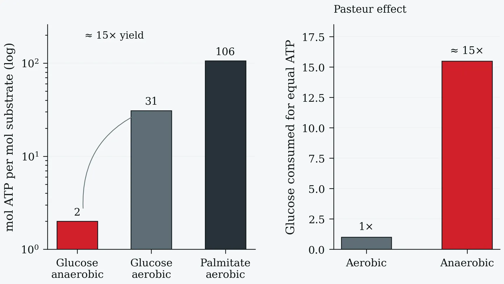
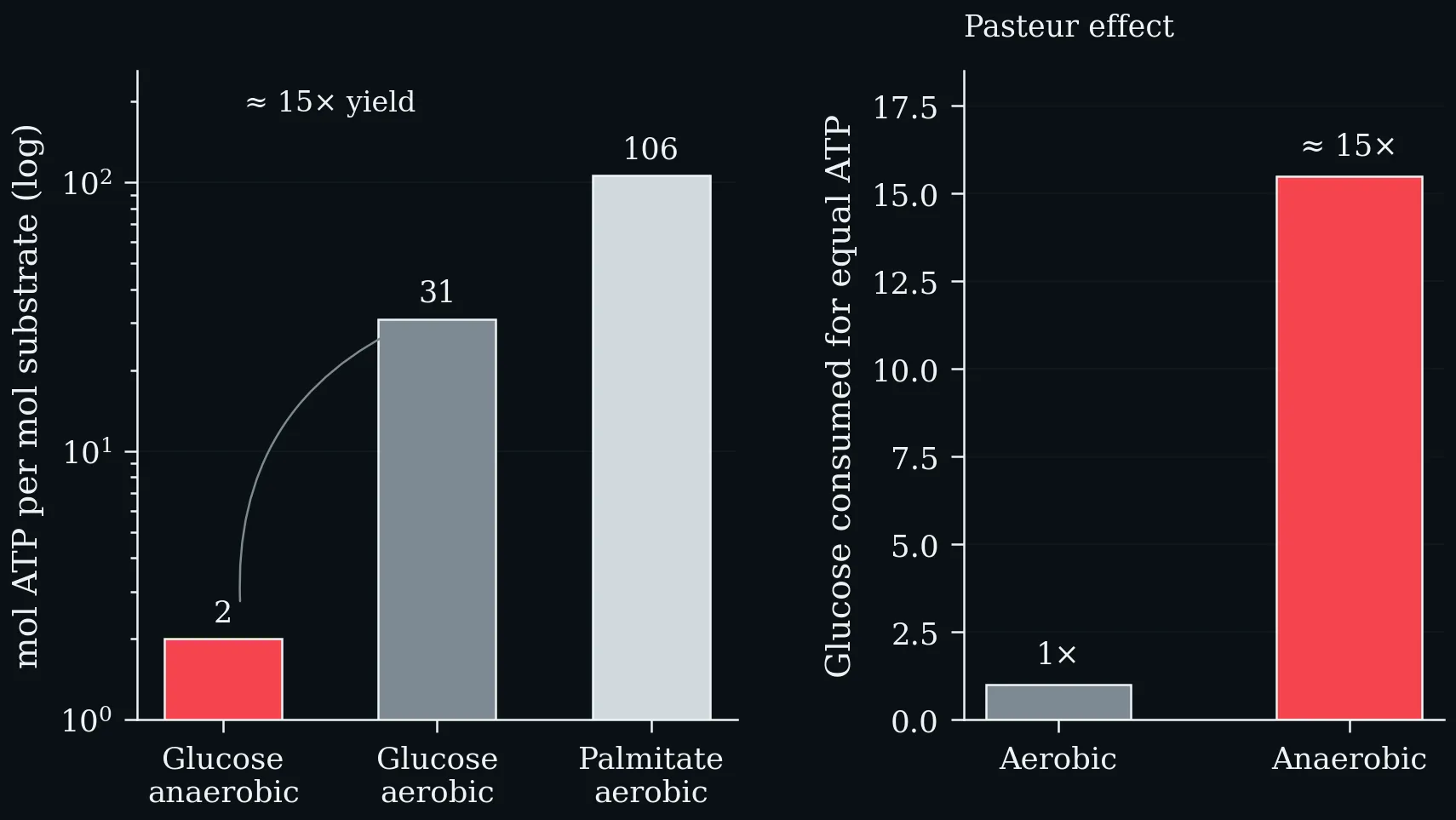
Left: ATP yield per mole of substrate on a logarithmic scale — 2 for anaerobic glycolysis, ~31 for the complete aerobic oxidation of glucose, and 106 for palmitate. Right: the direct consequence, the Pasteur effect. Because the anaerobic yield is roughly 15-fold lower, about 15× as much glucose must be consumed anaerobically to liberate the same amount of ATP (Nelson & Cox 2013).
Glycolytic flux is regulated to keep ATP nearly constant, via allosteric control of hexokinase, PFK-1 and pyruvate kinase, and second-to-second metabolite fluctuations; on a slower scale by glucagon, epinephrine and insulin, and by enzyme gene expression.
The Warburg effect. Warburg observed (1928) that tumours of nearly all types carry out glycolysis at a much higher rate than normal tissue, even when oxygen is available (Nelson & Cox 2013):
- Tumour cells rely on glycolysis for much ATP; the low yield (2 vs ~30 ATP/glucose) forces high glucose uptake.
- The hypoxia-inducible factor HIF-1 stimulates ≥8 glycolytic enzymes and the transporters GLUT1/GLUT3 when O₂ is limited.
- Three hexokinase inhibitors show chemotherapeutic promise: 2-deoxyglucose, lonidamine, 3-bromopyruvate.
- PET with ¹⁸F-labelled 2-fluoro-2-deoxyglucose (FdG) detects tumours by their high glucose uptake.
2.4 Glucose Uptake, GLUT4 and Type 1 Diabetes
Glucose uptake is mediated by the GLUT family. Hepatocyte (GLUT1/2) and neuronal (GLUT3) transporters are constitutively present; the main transporter of skeletal muscle, cardiac muscle and adipose tissue (GLUT4) is sequestered in intracellular vesicles and moves to the membrane only in response to an insulin signal (Nelson & Cox 2013).
In type 1 diabetes, too few β cells means insufficient insulin to trigger GLUT4-mediated uptake. Glucose accumulates (hyperglycaemia); muscle and fat instead burn fatty acids, and hepatic acetyl-CoA is converted to ketone bodies (acetoacetate, β-hydroxybutyrate). Overproduction lowers blood pH — ketoacidosis, a life-threatening condition (Nelson & Cox 2013).
Exercise relevance. Exercise stimulates GLUT4 translocation via AMPK and calcium signalling, independent of insulin (Richter & Hargreaves 2013) — of particular clinical importance in diabetes. The Lehninger Fig. 14-10 cascade (insulin defect → blocked PI-3K/PKB → no GLUT4 insertion → blocked glycolysis and PPP → impaired TCA/ETC → triacylglycerol mobilisation → ketoacidosis) makes each step of this failure explicit (Nelson & Cox 2013).
2.5 Fermentation and the Cori Cycle
Fermentation is the anaerobic degradation of glucose (or other nutrients) to obtain ATP; it neither consumes O₂ nor changes net NAD⁺/NADH. In lactic-acid fermentation, NADH from glycolysis is reoxidised to NAD⁺ by reducing pyruvate to lactate (lactate dehydrogenase):
Pyruvate + NADH + H⁺ → L-Lactate + NAD⁺ (ΔG′° = −25.1 kJ/mol)
In ethanol fermentation, pyruvate is decarboxylated to acetaldehyde (pyruvate decarboxylase; TPP, Mg²⁺) then reduced to ethanol by alcohol dehydrogenase using NADH (Nelson & Cox 2013).
In exercise, during a sprint the blood cannot deliver O₂ fast enough; muscle ferments stored glycogen to lactate. Lactate is later reconverted to glucose by hepatic gluconeogenesis during recovery, when the excess O₂ consumed repays the oxygen debt. The muscle-lactate → liver-glucose loop is the Cori cycle (Carl and Gerty Cori). A trained athlete recovers from a 100 m sprint in ≤30 min; an alligator may need many hours (Nelson & Cox 2013).
2.6 Gluconeogenesis
Some tissues depend almost entirely on glucose — the brain alone needs ~120 g/day. Between meals, during fasting, or after vigorous exercise, glycogen is depleted, and glucose must be made from non-carbohydrate precursors by gluconeogenesis (Nelson & Cox 2013):
- Occurs in all animals, plants, fungi and microorganisms; mainly in the liver (also renal cortex, small-intestinal epithelium).
- Important precursors are three-carbon compounds — lactate, pyruvate, glycerol — and glucogenic amino acids.
- After exercise, muscle lactate returns to the liver, is converted to glucose, and returns to muscle as glycogen (Cori cycle).
Not simply reverse glycolysis. Seven of ten reactions reverse glycolysis, but three irreversible glycolytic steps are bypassed (Nelson & Cox 2013):
- Pyruvate → PEP — pyruvate carboxylase (mitochondrial; biotin, ATP → oxaloacetate) then PEP carboxykinase (GTP).
- Fructose 1,6-bisphosphate → fructose 6-phosphate — fructose 1,6-bisphosphatase (Mg²⁺; ΔG′° = −16.3 kJ/mol).
- Glucose 6-phosphate → glucose — glucose 6-phosphatase (ER lumen of hepatocytes, renal and intestinal cells; ΔG′° = −13.8 kJ/mol).
Gluconeogenesis is energetically expensive:
2 Pyruvate + 4ATP + 2GTP + 2NADH + 2H⁺ + 4H₂O → Glucose + 4ADP + 2GDP + 6Pᵢ + 2NAD⁺
Six high-energy phosphates per glucose (vs 2 ATP for glycolysis). Glycolysis and gluconeogenesis are reciprocally regulated to prevent futile cycling (Nelson & Cox 2013).
2.7 Pentose Phosphate Pathway
The pentose phosphate pathway (phosphogluconate / hexose monophosphate pathway) is an alternative catabolic fate of glucose 6-phosphate with two major functions (Nelson & Cox 2013):
- NADPH production — for fatty-acid, cholesterol and steroid synthesis (liver, adipose, adrenal, gonad) and for maintaining reduced glutathione against oxidative damage (erythrocytes, lens, cornea).
- Ribose 5-phosphate production — for RNA, DNA and coenzyme synthesis in rapidly dividing cells (marrow, skin, intestinal mucosa, tumours).
Oxidative phase: glucose 6-phosphate dehydrogenase (G6PD) reduces NADP⁺ to NADPH; after lactonase and 6-phosphogluconate dehydrogenase, a second NADPH forms and ribulose 5-phosphate is produced, isomerised to ribose 5-phosphate.
Glucose 6-phosphate + 2NADP⁺ + H₂O → ribose 5-phosphate + CO₂ + 2NADPH + 2H⁺
Non-oxidative phase: transketolase (TPP cofactor) and transaldolase interconvert three-, four-, five-, six- and seven-carbon sugars, reversibly converting six pentose phosphates to five hexose phosphates. Whether glucose 6-phosphate enters glycolysis or the PPP depends on the cytosolic NADP⁺/NADPH ratio: rising NADP⁺ allosterically stimulates G6PD and diverts flux to the PPP (Nelson & Cox 2013).
One-Minute-Paper Topics
A One-Minute-Paper (OMP) is a short, focused prompt that students answer in ~60 seconds at the end of a session to consolidate learning, surface misconceptions, and provide formative feedback. When answering, be concise, specific, and use terminology from today’s session.
- Describe the four major fates of glucose and the three catabolic fates of pyruvate, and the conditions that favour each.
- Explain the Warburg effect in one sentence and name one context in which high aerobic glycolysis also occurs in healthy tissue.
- Why is GLUT4 special among glucose transporters, and how does exercise recruit it independently of insulin?
- What is gluconeogenesis, why is it not simply reverse glycolysis, and why does it matter during prolonged exercise?
- Briefly explain the two functions of the pentose phosphate pathway and when a cell diverts glucose 6-phosphate into it.
Key References
- Nelson DL, Cox MM. Lehninger Principles of Biochemistry, 6th edn. W.H. Freeman and Company, 2013.
- Richter EA, Hargreaves M. Exercise, GLUT4, and skeletal muscle glucose uptake. Physiol Rev 93 (3), 2013: 993–1017.
3 Substrate Utilization and Hormonal Control
Learning objectives:
- Carbohydrate, fat and protein as exercise substrates, and their relative contributions
- FATmax and the carbohydrate-to-fat crossover as intensity rises
- Hormonal control of metabolism: catecholamines, insulin/glucagon, cortisol/growth hormone
3.1 Carbohydrate Metabolism
Carbohydrates are the primary fuel during moderate-to-high-intensity exercise; muscle glycogen and blood glucose are the main substrates (Coggan & Coyle 1991). Glycogen depletion rate rises with intensity — above ~70 % V̇O₂max, glycogen use increases exponentially, and depletion produces the fatigue known as “hitting the wall” in endurance events (Bergström et al. 1967). Glucose uptake is enhanced during exercise through both insulin-dependent and insulin-independent mechanisms, the latter via AMPK- and calcium-driven GLUT4 translocation (Richter & Hargreaves 2013).
3.2 Fat Metabolism
Fat oxidation supplies energy during low-to-moderate intensity exercise and grows in importance as duration extends. Fatty acids enter mitochondria via the carnitine shuttle before beta-oxidation (Jeukendrup 2002). The maximal rate of fat oxidation (FATmax) typically occurs at ~45–65 % V̇O₂max and shifts with training status; endurance training raises fat-oxidation capacity by increasing mitochondrial density, oxidative enzyme activity and capillary density (Achten et al. 2002).
3.3 Protein Metabolism
Proteins contribute minimally to most exercise (typically 5–15 % of total energy) but become more significant during prolonged exercise when glycogen is depleted. Amino acids, particularly branched-chain amino acids (BCAAs), can be oxidised for energy or used for gluconeogenesis (Wagenmakers 1998).
3.4 Substrate Utilization at Different Exercise Intensities
The crossover from fat to carbohydrate as exercise intensity rises. At low intensity the CHO:fat ratio is about 30:70, near 50:50 at moderate intensity, and 85:15 at high intensity. FAT_max — the intensity at which absolute fat oxidation peaks — lies at roughly 45–65 % V̇O₂max, well below the intensity at which carbohydrate begins to dominate the mixture.
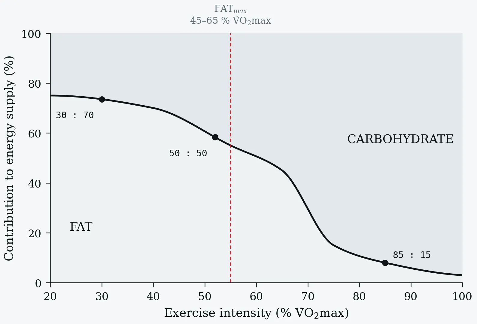
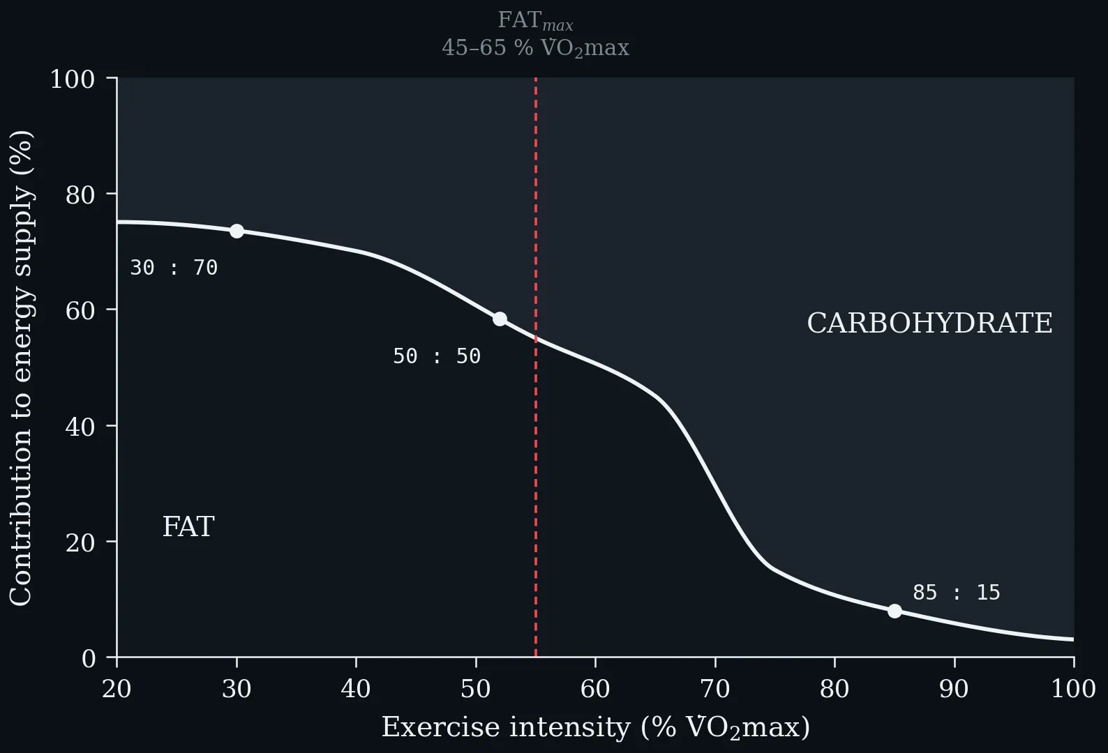
The crossover from fat to carbohydrate as exercise intensity rises. At low intensity the CHO:fat ratio is about 30:70, near 50:50 at moderate intensity, and 85:15 at high intensity. FAT_max — the intensity at which absolute fat oxidation peaks — lies at roughly 45–65 % V̇O₂max, well below the intensity at which carbohydrate begins to dominate the mixture.
Exercise triggers profound hormonal responses that coordinate these metabolic adjustments to meet energy demands and maintain homeostasis (Galbo 1983).
3.5 Catecholamines (Epinephrine and Norepinephrine)
Catecholamine secretion increases proportionally with exercise intensity. These hormones promote glycogenolysis and lipolysis through β-adrenergic receptor stimulation, increasing both glucose and free-fatty-acid availability, and they enhance cardiac output and redirect blood flow to working muscles (Kjaer 1989).
3.6 Insulin and Glucagon
During exercise, insulin falls while glucagon rises, promoting hepatic glucose production to maintain blood-glucose homeostasis. This reciprocal regulation ensures adequate glucose delivery to working muscle and the central nervous system (Wasserman 2009).
3.7 Cortisol and Growth Hormone
Both increase during prolonged exercise. Cortisol stimulates gluconeogenesis and protein catabolism; growth hormone promotes lipolysis and exerts anabolic effects during recovery. Their elevation becomes particularly significant during exercise exceeding 60 minutes (Viru & Viru 2004).
One-Minute-Paper Topics
A One-Minute-Paper (OMP) is a short, focused prompt that students answer in ~60 seconds at the end of a session to consolidate learning, surface misconceptions, and provide formative feedback. When answering, be concise, specific, and use terminology from today’s session.
- How does substrate utilisation shift from rest to high-intensity exercise, and what is FATmax?
- Describe one action of catecholamines on exercise metabolism.
- Contrast the metabolic effects of insulin and glucagon during and after exercise.
- How do cortisol and growth hormone contribute to substrate mobilisation during prolonged exercise?
Key References
- Achten J, Gleeson M, Jeukendrup AE. Determination of the exercise intensity that elicits maximal fat oxidation. Med Sci Sports Exerc 34 (1), 2002: 92–97.
- Bergström J, Hermansen L, Hultman E, Saltin B. Diet, muscle glycogen and physical performance. Acta Physiol Scand 71 (2), 1967: 140–150.
- Coggan AR, Coyle EF. Carbohydrate ingestion during prolonged exercise: effects on metabolism and performance. Exerc Sport Sci Rev 19, 1991: 1–40.
- Galbo H. Hormonal and Metabolic Adaptation to Exercise. Thieme Medical Publishers, 1983.
- Jeukendrup AE. Regulation of fat metabolism in skeletal muscle. Ann N Y Acad Sci 967, 2002: 217–235.
- Kjaer M. Epinephrine and some other hormonal responses to exercise in man. Int J Sports Med 10 (1), 1989: 2–15.
- Richter EA, Hargreaves M. Exercise, GLUT4, and skeletal muscle glucose uptake. Physiol Rev 93 (3), 2013: 993–1017.
- Viru A, Viru M. Cortisol — essential adaptation hormone in exercise. Int J Sports Med 25 (6), 2004: 461–464.
- Wagenmakers AJ. Protein and amino acid metabolism in human muscle. Adv Exp Med Biol 441, 1998: 307–319.
- Wasserman DH. Four grams of glucose. Am J Physiol Endocrinol Metab 296 (1), 2009: E11–E21.
4 Lactate Metabolism and the Lactate Threshold
Learning objectives:
- Why lactate is now viewed as a fuel and signalling molecule rather than a waste product
- Production (glycolysis, NAD⁺ regeneration, glutamine) and clearance (PDH, gluconeogenesis/Cori)
- MCT transport and the lactate shuttle, GPR81 signalling and histone lactylation, and the lactate threshold
4.1 The Lactate Paradigm Shift
Since its discovery in 1780, lactate was wrongly characterised as a harmful by-product produced only under hypoxia. Research over recent decades has revolutionised this view: lactate is a signalling molecule, an energy substrate, and a key metabolic regulator, produced and used even under fully aerobic conditions (Li et al. 2022). Understanding lactate homeostasis — production, clearance and regulation — is central to exercise physiology, cancer metabolism and inflammation.
4.2 Lactate Production Pathways
Lactate production rises when demand for O₂ and ATP exceeds supply — strenuous exercise, infection/inflammation, hypoxia, cancer.
Primary pathway — glycolysis. Glucose is broken to two pyruvate. Under aerobic conditions pyruvate enters mitochondria → acetyl-CoA → TCA; under high demand or hypoxia the TCA cycle is insufficient and pyruvate is reduced directly to lactate by lactate dehydrogenase (LDH) (Nelson & Cox 2013).
The critical role of NAD⁺ regeneration. Lactate production is not just about making a metabolite — it regenerates NAD⁺. Glycolysis requires NAD⁺; converting pyruvate to lactate reoxidises NADH to NAD⁺, letting glycolysis continue producing ATP when mitochondria cannot keep up. Without it, glycolysis would halt from NAD⁺ depletion.
Secondary pathway — glutamine catabolism (important in cancer): glutamine → glutamate (glutaminase, often upregulated by c-Myc) → α-ketoglutarate → TCA; glutamine-derived carbon becomes oxaloacetate → malate, which exits the mitochondria and is converted by malic enzyme (ME1) to NADPH and pyruvate — a secondary lactate source. This serves dual purposes: NADPH production and lactate production (Li et al. 2022).
4.3 Lactate Clearance Mechanisms
Accumulating lactate can cause lactic acidosis, so it must be rapidly metabolised.
Irreversible — the PDH pathway. Lactate is oxidised back to pyruvate; pyruvate dehydrogenase (PDH) converts pyruvate to acetyl-CoA, which enters the TCA cycle for complete oxidation. This is a one-way street: acetyl-CoA cannot revert to a three-carbon unit. PDH is regulated by E1α phosphorylation (phosphorylated = inactive) and NADH (high NADH inhibits). When mitochondrial respiration is impaired, PDH activity falls and circulating lactate rises — why lactate is a valuable clinical marker (Li et al. 2022).
Reversible — gluconeogenesis and the Cori cycle. In liver (and muscle), lactate → pyruvate → glucose, which is released to blood — maintaining glucose during fasting or prolonged exercise and recycling lactate. Muscle lactate → hepatic glucose → back to muscle is the Cori cycle (Brooks 2009).
4.4 Lactate Transport and Regulation
Monocarboxylate transporters (MCTs). Lactate crosses membranes via MCTs (SLC16 family; 14 identified, MCT1–4 most important). They catalyse proton-coupled, bidirectional transport (lactate:H⁺ 1:1 symport).
- MCT1 (homeostatic): widely distributed, high affinity, basal lactate maintenance along the gradient.
- MCT4 (high-capacity): low affinity, high capacity, expressed in highly glycolytic tissue, tumour cells and white (Type II) fibres. High MCT1/2/4 expression is associated with cancer progression.
The lactate shuttle. Synergistic MCT1–4 activity shuttles lactate from glycolytic producers to oxidative consumers: in muscle, Type II → Type I fibres; in tumours, hypoxic → normoxic cells; in brain, astrocytes → neurons.
GPR81 — the lactate receptor. Lactate signals through G protein-coupled receptor 81 (GPR81) (highest in adipose; also kidney, muscle, CNS, heart), mediating energy metabolism, lipolysis regulation, neuronal protection and inflammatory regulation — making lactate a true signalling molecule (Li et al. 2022).
4.5 Cellular Lactate Metabolism and Lactylation
Intracellular lactate arises from glycolysis (LDH) and the glutamine/ME1 route; within mitochondria it can be reconverted to pyruvate (PDH → acetyl-CoA → TCA), connecting to cholesterol and lipid synthesis, or cleared reversibly via gluconeogenesis (lactate → pyruvate → glucose, mainly in liver).
Protein lactylation. One of the most revolutionary recent discoveries: lactate is a substrate for protein lactylation, a post-translational modification with epigenetic consequences (Li et al. 2022):
- Lactyl-CoA forms from lactate, serving as the modification substrate.
- Histone lactylation (Kla) modifies lysine residues; p300 is the “writer”, HDACs the “erasers”.
- Non-histone proteins can also be lactylated, altering their function.
- Lactylated histones control genes of inflammation, tumour development, immune responses and metabolism (e.g. Arg1, YTHDF2).
Lactate is therefore an epigenetic regulator, not merely a metabolite.
4.6 Lactate Homeostasis and the Lactate Threshold
Lactate homeostasis is a balance:
[Lactate]_steady-state = Production − Clearance
where Production = glycolysis + glutamine metabolism + MCT import, and Clearance = PDH oxidation + gluconeogenesis + MCT export + lactylation. Key determinants: glycolytic flux, PDH activity, MCT expression, O₂ availability, and metabolic demand.
The lactate threshold is the exercise intensity at which blood lactate begins to accumulate exponentially — typically 50–60 % V̇O₂max in untrained individuals and 70–80 % V̇O₂max in endurance athletes. It is a strong, highly trainable predictor of endurance performance (Faude et al. 2009). Rather than accumulating passively, lactate is continuously produced and cleared even at rest, and during exercise is oxidised in the same fibre, shuttled to oxidative fibres, or converted to glucose in the liver (Brooks 2009).
Summary — the paradigm shift. Old view (pre-1980s): a waste product of anaerobic metabolism causing fatigue. Current view: an essential metabolite and signalling molecule, produced both aerobically and anaerobically, functioning as (1) an energy substrate (often preferred over glucose), (2) a redox buffer (NAD⁺/NADH), (3) a signalling molecule (GPR81), and (4) an epigenetic regulator (lactylation) (Li et al. 2022; Brooks 2018).
One-Minute-Paper Topics
A One-Minute-Paper (OMP) is a short, focused prompt that students answer in ~60 seconds at the end of a session to consolidate learning, surface misconceptions, and provide formative feedback. When answering, be concise, specific, and use terminology from today’s session.
- Why is lactate now considered a fuel and signalling molecule rather than a waste product?
- Explain, in one sentence each, the role of NAD⁺ regeneration in lactate production and the lactate-shuttle concept.
- Contrast irreversible (PDH) and reversible (gluconeogenesis/Cori) lactate clearance.
- What is the role of MCT transporters in lactate exchange between tissues, and how do MCT1 and MCT4 differ?
- Define protein lactylation and explain its potential epigenetic relevance; how does GPR81 signalling extend lactate’s role?
Key References
- Brooks GA. Cell-cell and intracellular lactate shuttles. J Physiol 587 (23), 2009: 5591–5600.
- Brooks GA. The science and translation of lactate shuttle theory. Cell Metab 27 (4), 2018: 757–785.
- Faude O, Kindermann W, Meyer T. Lactate threshold concepts: how valid are they? Sports Med 39 (6), 2009: 469–490.
- Li X, Yang Y, Zhang B, et al. Lactate metabolism in human health and disease. Signal Transduct Target Ther 7 (1), 2022: 305.
- Nelson DL, Cox MM. Lehninger Principles of Biochemistry, 6th edn. W.H. Freeman and Company, 2013.
5 Fibers, Adaptations, Recovery and Clinical Applications
Learning objectives:
- Contractile and metabolic properties of Type I, IIa and IIx fibres
- Endurance vs resistance adaptations, the opposing AMPK and mTOR pathways, and ROS as adaptive signals (hormesis)
- Recovery metabolism (EPOC, glycogen resynthesis, MPS) and exercise as medicine
5.1 Muscle Fiber Types and Metabolic Characteristics
Skeletal muscle contains heterogeneous fibre types with distinct metabolic and contractile properties (Schiaffino & Reggiani 2011).
Type I (slow-twitch, oxidative): high mitochondrial density and oxidative enzyme content, rich capillary supply and myoglobin (red), high fatigue resistance, lower force and contraction speed, primary reliance on fat oxidation.
Type II (fast-twitch): subdivided into Type IIa (fast oxidative-glycolytic, intermediate properties) and Type IIx (fast glycolytic — highest glycolytic and force capacity, lowest oxidative capacity, fatigues rapidly) (Bottinelli & Reggiani 2000).
Contractile and metabolic characteristics of the three skeletal-muscle fiber types.
| Characteristic | Type I | Type IIa | Type IIx |
|---|---|---|---|
| Contraction Speed | Slow | Fast | Very Fast |
| Force Production | Low | High | Very High |
| Mitochondrial Density | High | Moderate | Low |
| Fatigue Resistance | High | Moderate | Low |
| Primary Metabolism | Oxidative | Oxid-Glycolytic | Glycolytic |
Contractile and metabolic characteristics of the three skeletal-muscle fiber types.
5.2 Biochemical Adaptations to Training
Endurance training enhances oxidative capacity and substrate utilisation (Holloszy 2008): mitochondrial biogenesis via PGC-1α; greater oxidative enzyme activity; increased capillary density; greater myoglobin; glycogen sparing at submaximal intensities; improved lactate clearance.
Resistance training stimulates muscle protein synthesis and neural adaptation (Phillips 2014): increased MPS via mTOR; enhanced PCr stores and creatine kinase activity; greater glycolytic enzyme content; Type II hypertrophy; satellite-cell activation.
Molecular signalling. AMPK is activated by cellular energy stress and drives glucose uptake, fat oxidation and mitochondrial biogenesis (Hardie 2011). mTOR integrates nutrients, growth factors and mechanical tension to regulate protein synthesis — activated by resistance loading, and transiently suppressed by acute endurance exercise (Bodine et al. 2001). PGC-1α is the master regulator of mitochondrial biogenesis, upregulated by endurance exercise via AMPK, calcium signalling and p38 MAPK (Puigserver et al. 1998). AMPK and mTOR thus represent broadly opposing adaptive axes for endurance vs resistance training.
5.3 Reactive Oxygen Species and Oxidative Stress
Exercise increases muscle O₂ consumption up to 100-fold, raising reactive oxygen species (ROS) production. While excessive ROS damages cellular components, moderate ROS serve as secondary messengers in exercise-induced adaptations — mitochondrial biogenesis, antioxidant enzyme expression and glucose uptake (Powers & Jackson 2008). The concept of hormesis holds that low-to-moderate oxidative stress stimulates adaptive responses that strengthen cellular defences (Ristow et al. 2009). Chronic training enhances endogenous antioxidant systems (SOD, catalase, glutathione peroxidase), improving the capacity to manage oxidative stress in subsequent bouts (Ji 2008).
5.4 Recovery Metabolism and Nutritional Biochemistry
EPOC. After exercise, O₂ consumption stays elevated to restore homeostasis — PCr resynthesis, lactate clearance, restoration of O₂ stores, elevated temperature, increased cardiac/respiratory work, catecholamine effects (Borsheim & Bahr 2003). Magnitude and duration scale with intensity and duration; high-intensity interval training produces greater EPOC than steady-state work of similar total work (LaForgia et al. 2006).
Glycogen resynthesis occurs in two phases: a rapid, insulin-independent phase (first 30–60 min, driven by exercise-induced GLUT4 translocation and glycogen synthase activity), then a slower insulin-dependent phase (Jentjens & Jeukendrup 2003). Maximal rates occur with ~1.0–1.2 g/kg/h carbohydrate; co-ingesting protein (0.2–0.4 g/kg) helps when carbohydrate is suboptimal (van Loon et al. 2000).
Muscle protein synthesis is stimulated for 24–48 h after (especially resistance) exercise (Burd et al. 2011). Post-exercise protein enhances MPS and reduces breakdown; leucine activates mTOR; optimal doses are ~20–40 g depending on body size and stimulus (Moore et al. 2009).
5.5 Clinical Applications and Exercise as Medicine
Exercise biochemistry underpins disease prevention and management (Pedersen & Saltin 2015).
Metabolic disease. Exercise improves insulin sensitivity via increased GLUT4 expression, better mitochondrial function and reduced intramuscular lipid — beneficial for preventing/managing type 2 diabetes (Hawley & Lessard 2008). It favourably modifies lipids (↑ HDL, ↓ triglycerides, improved LDL particle size) through enhanced lipoprotein lipase activity and altered hepatic lipid metabolism (Durstine et al. 2001).
Cardiovascular health. Training improves endothelial function, arterial compliance and cardiac efficiency; enhanced nitric-oxide bioavailability improves vasodilation and lowers vascular oxidative stress (Green et al. 2004). Cardiac adaptations (↑ stroke volume, improved diastolic filling, enhanced contractility) reduce cardiac workload at a given absolute load (Blomqvist & Saltin 1983).
Ageing and sarcopenia. Resistance training counteracts age-related muscle loss by stimulating MPS and satellite-cell activation; with adequate protein it maintains muscle mass and function in older adults (Fiatarone et al. 1994). Exercise also preserves mitochondrial function with age, reducing damaged-mitochondria accumulation and frailty (Short et al. 2005).
5.6 Key Terms Related to Physical Activity and Exercise Prescription
Physical activity: any bodily movement produced by skeletal-muscle contraction that increases energy expenditure above basal level — incidental (activities of daily living) and intentional (e.g. non-structured activity such as walking the dog).
Exercise: planned, structured physical activity performed with the explicit intent of improving or maintaining components of physical fitness (aerobic capacity, strength, power, endurance, body composition, balance, coordination, flexibility) and/or a specific health benefit.
Sport: rule-governed, structured, competitive physical activity involving gross motor movement.
Cardiorespiratory fitness: the integrated ability of the circulatory and respiratory systems to supply O₂ to skeletal-muscle mitochondria; often expressed as V̇O₂max or estimated from work rate or non-exercise algorithms. Cardiorespiratory exercise requires both systems to support sustained metabolism (preferred over “aerobic” or “endurance” exercise).
Metabolic threshold: an intensity producing an apparent change in metabolism (first and second metabolic thresholds).
Body composition: relative proportions of fat mass and fat-free mass. Muscle strength (force in a task), muscle power (force × velocity, or rate of work), muscle endurance (sustained force), muscle fitness (the global term).
Resistance exercise: exercise requiring muscles to exert force against resistance (body weight, bands, free weights, machines) until repetitions/duration are limited by neuromuscular fatigue.
Flexibility: the ability to move a joint through its maximal unrestricted range of motion without pain. Perception of effort: the cognitive feeling of effort integrating signals from peripheral muscles/joints, the cardiopulmonary system and the CNS.
5.7 Conclusion
Exercise biochemistry reveals the molecular and metabolic adaptations enabling human performance and health — from immediate energy provision (ATP-PCr) to long-term adaptations in mitochondrial density and protein synthesis. Understanding these principles supports evidence-based training, nutrition and therapeutic intervention, and reinforces exercise as a powerful stimulus for positive adaptation and a cornerstone of preventive medicine.
One-Minute-Paper Topics
A One-Minute-Paper (OMP) is a short, focused prompt that students answer in ~60 seconds at the end of a session to consolidate learning, surface misconceptions, and provide formative feedback. When answering, be concise, specific, and use terminology from today’s session.
- Compare Type I and Type II muscle fibres in terms of biochemical and metabolic properties.
- Describe how AMPK and mTOR signalling represent opposing adaptive pathways to endurance vs resistance training.
- Why would suppressing exercise-induced ROS entirely be expected to blunt training adaptations?
- What is EPOC, and which biochemical processes drive it after high-intensity exercise?
- Give one clinical application of exercise biochemistry relevant to post-infection recovery.
Key References
- Blomqvist CG, Saltin B. Cardiovascular adaptations to physical training. Annu Rev Physiol 45, 1983: 169–189.
- Bodine SC, Stitt TN, Gonzalez M, et al. Akt/mTOR pathway is a crucial regulator of skeletal muscle hypertrophy. Nat Cell Biol 3 (11), 2001: 1014–1019.
- Borsheim E, Bahr R. Effect of exercise intensity, duration and mode on post-exercise oxygen consumption. Sports Med 33 (14), 2003: 1037–1060.
- Bottinelli R, Reggiani C. Human skeletal muscle fibres: molecular and functional diversity. Prog Biophys Mol Biol 73 (2-4), 2000: 195–262.
- Burd NA, West DW, Moore DR, et al. Enhanced amino acid sensitivity of myofibrillar protein synthesis persists for up to 24 h after resistance exercise in young men. J Nutr 141 (4), 2011: 568–573.
- Durstine JL, Grandjean PW, Davis PG, et al. Blood lipid and lipoprotein adaptations to exercise. Sports Med 31 (15), 2001: 1033–1062.
- Fiatarone MA, O’Neill EF, Ryan ND, et al. Exercise training and nutritional supplementation for physical frailty in very elderly people. N Engl J Med 330 (25), 1994: 1769–1775.
- Green DJ, Maiorana A, O’Driscoll G, Taylor R. Effect of exercise training on endothelium-derived nitric oxide function in humans. J Physiol 561 (1), 2004: 1–25.
- Hardie DG. AMP-activated protein kinase: an energy sensor that regulates all aspects of cell function. Genes Dev 25 (18), 2011: 1895–1908.
- Hawley JA, Lessard SJ. Exercise training-induced improvements in insulin action. Acta Physiol 192 (1), 2008: 127–135.
- Holloszy JO. Regulation by exercise of skeletal muscle content of mitochondria and GLUT4. J Physiol Pharmacol 59 (Suppl 7), 2008: 5–18.
- Jentjens R, Jeukendrup AE. Determinants of post-exercise glycogen synthesis during short-term recovery. Sports Med 33 (2), 2003: 117–144.
- Ji LL. Modulation of skeletal muscle antioxidant defense by exercise: role of redox signaling. Free Radic Biol Med 44 (2), 2008: 142–152.
- LaForgia J, Withers RT, Gore CJ. Effects of exercise intensity and duration on the excess post-exercise oxygen consumption. J Sports Sci 24 (12), 2006: 1247–1264.
- Moore DR, Robinson MJ, Fry JL, et al. Ingested protein dose response of muscle and albumin protein synthesis after resistance exercise in young men. Am J Clin Nutr 89 (1), 2009: 161–168.
- Pedersen BK, Saltin B. Exercise as medicine — evidence for prescribing exercise as therapy in 26 different chronic diseases. Scand J Med Sci Sports 25 (Suppl 3), 2015: 1–72.
- Phillips SM. A brief review of critical processes in exercise-induced muscular hypertrophy. Sports Med 44 (Suppl 1), 2014: 71–77.
- Powers SK, Jackson MJ. Exercise-induced oxidative stress: cellular mechanisms and impact on muscle force production. Physiol Rev 88 (4), 2008: 1243–1276.
- Puigserver P, Wu Z, Park CW, et al. A cold-inducible coactivator of nuclear receptors linked to adaptive thermogenesis. Cell 92 (6), 1998: 829–839.
- Ristow M, Zarse K, Oberbach A, et al. Antioxidants prevent health-promoting effects of physical exercise in humans. Proc Natl Acad Sci USA 106 (21), 2009: 8665–8670.
- Schiaffino S, Reggiani C. Fiber types in mammalian skeletal muscles. Physiol Rev 91 (4), 2011: 1447–1531.
- Short KR, Bigelow ML, Kahl J, et al. Decline in skeletal muscle mitochondrial function with aging in humans. Proc Natl Acad Sci USA 102 (15), 2005: 5618–5623.
- van Loon LJ, Saris WH, Kruijshoop M, Wagenmakers AJ. Maximizing postexercise muscle glycogen synthesis. Am J Clin Nutr 72 (1), 2000: 106–111.
References
- Achten J, Gleeson M, Jeukendrup AE. Determination of the exercise intensity that elicits maximal fat oxidation. Med Sci Sports Exerc 34 (1), 2002: 92–97.
- Berg JM, Tymoczko JL, Stryer L. Biochemistry, 5th edn. W.H. Freeman and Company, 2002.
- Bergström J, Hermansen L, Hultman E, Saltin B. Diet, muscle glycogen and physical performance. Acta Physiol Scand 71 (2), 1967: 140–150.
- Bishop DJ, Beck B, Biddle SJH, et al. Physical activity and exercise intensity terminology. J Sci Med Sport 28, 2025: 980–991.
- Blomqvist CG, Saltin B. Cardiovascular adaptations to physical training. Annu Rev Physiol 45, 1983: 169–189.
- Bodine SC, Stitt TN, Gonzalez M, et al. Akt/mTOR pathway is a crucial regulator of skeletal muscle hypertrophy. Nat Cell Biol 3 (11), 2001: 1014–1019.
- Borsheim E, Bahr R. Effect of exercise intensity, duration and mode on post-exercise oxygen consumption. Sports Med 33 (14), 2003: 1037–1060.
- Bottinelli R, Reggiani C. Human skeletal muscle fibres: molecular and functional diversity. Prog Biophys Mol Biol 73 (2-4), 2000: 195–262.
- Brooks GA. Cell-cell and intracellular lactate shuttles. J Physiol 587 (23), 2009: 5591–5600.
- Brooks GA. The science and translation of lactate shuttle theory. Cell Metab 27 (4), 2018: 757–785.
- Brooks GA, Fahey TD, Baldwin KM. Exercise Physiology: Human Bioenergetics and Its Applications, 4th edn. McGraw-Hill, 2005.
- Burd NA, West DW, Moore DR, et al. Enhanced amino acid sensitivity of myofibrillar protein synthesis persists for up to 24 h after resistance exercise in young men. J Nutr 141 (4), 2011: 568–573.
- Coggan AR, Coyle EF. Carbohydrate ingestion during prolonged exercise: effects on metabolism and performance. Exerc Sport Sci Rev 19, 1991: 1–40.
- Durstine JL, Grandjean PW, Davis PG, et al. Blood lipid and lipoprotein adaptations to exercise. Sports Med 31 (15), 2001: 1033–1062.
- Faude O, Kindermann W, Meyer T. Lactate threshold concepts: how valid are they? Sports Med 39 (6), 2009: 469–490.
- Fiatarone MA, O’Neill EF, Ryan ND, et al. Exercise training and nutritional supplementation for physical frailty in very elderly people. N Engl J Med 330 (25), 1994: 1769–1775.
- Galbo H. Hormonal and Metabolic Adaptation to Exercise. Thieme Medical Publishers, 1983.
- Green DJ, Maiorana A, O’Driscoll G, Taylor R. Effect of exercise training on endothelium-derived nitric oxide function in humans. J Physiol 561 (1), 2004: 1–25.
- Greenhaff PL. The creatine-phosphocreatine system: there’s more than one song in its repertoire. J Physiol 537 (3), 2001: 657.
- Hardie DG. AMP-activated protein kinase: an energy sensor that regulates all aspects of cell function. Genes Dev 25 (18), 2011: 1895–1908.
- Harris RC, Edwards RH, Hultman E, et al. The time course of phosphorylcreatine resynthesis during recovery of the quadriceps muscle in man. Pflügers Arch 367 (2), 1976: 137–142.
- Hawley JA, Hargreaves M, Joyner MJ, Zierath JR. Integrative biology of exercise. Cell 159 (4), 2014: 738–749.
- Hawley JA, Lessard SJ. Exercise training-induced improvements in insulin action. Acta Physiol 192 (1), 2008: 127–135.
- Holloszy JO. Regulation by exercise of skeletal muscle content of mitochondria and GLUT4. J Physiol Pharmacol 59 (Suppl 7), 2008: 5–18.
- Holloszy JO, Coyle EF. Adaptations of skeletal muscle to endurance exercise and their metabolic consequences. J Appl Physiol 56 (4), 1984: 831–838.
- Jentjens R, Jeukendrup AE. Determinants of post-exercise glycogen synthesis during short-term recovery. Sports Med 33 (2), 2003: 117–144.
- Jeukendrup AE. Regulation of fat metabolism in skeletal muscle. Ann N Y Acad Sci 967, 2002: 217–235.
- Ji LL. Modulation of skeletal muscle antioxidant defense by exercise: role of redox signaling. Free Radic Biol Med 44 (2), 2008: 142–152.
- Kjaer M. Epinephrine and some other hormonal responses to exercise in man. Int J Sports Med 10 (1), 1989: 2–15.
- LaForgia J, Withers RT, Gore CJ. Effects of exercise intensity and duration on the excess post-exercise oxygen consumption. J Sports Sci 24 (12), 2006: 1247–1264.
- Li X, Yang Y, Zhang B, et al. Lactate metabolism in human health and disease. Signal Transduct Target Ther 7 (1), 2022: 305.
- Moore DR, Robinson MJ, Fry JL, et al. Ingested protein dose response of muscle and albumin protein synthesis after resistance exercise in young men. Am J Clin Nutr 89 (1), 2009: 161–168.
- Nelson DL, Cox MM. Lehninger Principles of Biochemistry, 6th edn. W.H. Freeman and Company, 2013.
- Pedersen BK, Saltin B. Exercise as medicine — evidence for prescribing exercise as therapy in 26 different chronic diseases. Scand J Med Sci Sports 25 (Suppl 3), 2015: 1–72.
- Phillips SM. A brief review of critical processes in exercise-induced muscular hypertrophy. Sports Med 44 (Suppl 1), 2014: 71–77.
- Powers SK, Jackson MJ. Exercise-induced oxidative stress: cellular mechanisms and impact on muscle force production. Physiol Rev 88 (4), 2008: 1243–1276.
- Puigserver P, Wu Z, Park CW, et al. A cold-inducible coactivator of nuclear receptors linked to adaptive thermogenesis. Cell 92 (6), 1998: 829–839.
- Richter EA, Hargreaves M. Exercise, GLUT4, and skeletal muscle glucose uptake. Physiol Rev 93 (3), 2013: 993–1017.
- Ristow M, Zarse K, Oberbach A, et al. Antioxidants prevent health-promoting effects of physical exercise in humans. Proc Natl Acad Sci USA 106 (21), 2009: 8665–8670.
- Robergs RA, Ghiasvand F, Parker D. Biochemistry of exercise-induced metabolic acidosis. Am J Physiol Regul Integr Comp Physiol 287 (3), 2004: R502–R516.
- Schiaffino S, Reggiani C. Fiber types in mammalian skeletal muscles. Physiol Rev 91 (4), 2011: 1447–1531.
- Short KR, Bigelow ML, Kahl J, et al. Decline in skeletal muscle mitochondrial function with aging in humans. Proc Natl Acad Sci USA 102 (15), 2005: 5618–5623.
- van Loon LJ, Saris WH, Kruijshoop M, Wagenmakers AJ. Maximizing postexercise muscle glycogen synthesis. Am J Clin Nutr 72 (1), 2000: 106–111.
- Viru A, Viru M. Cortisol — essential adaptation hormone in exercise. Int J Sports Med 25 (6), 2004: 461–464.
- Wagenmakers AJ. Protein and amino acid metabolism in human muscle. Adv Exp Med Biol 441, 1998: 307–319.
- Wasserman DH. Four grams of glucose. Am J Physiol Endocrinol Metab 296 (1), 2009: E11–E21.